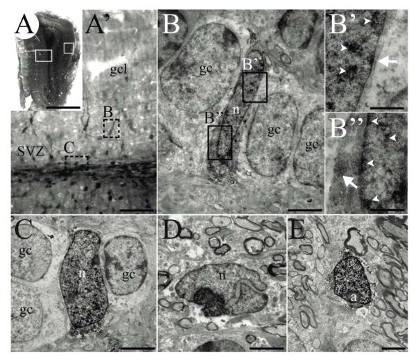
FIGURE 4. AORGs resemble immature glia ultrastructurally.
(A) Photomontage of a coronal section of the adult OB from an hGFAP-eGFP transgenic mouse in which expression of GFP is reported via immunoperoxidase staining. White boxed areas (left, right) indicate approximate regions from which images shown in A’ and E were obtained, respectively. (A’) Higher magnification image of left boxed area in A, showing deep layers of the OB, granule cell layer, and the anterior extension of the RMS. Note that the DAB precipitate reveals cells of different intensity in the granule cell layer and SVZ. Dashed boxes indicate areas shown in B and C. (B, B’, and B”) An example of an AORG, found between mature granule cells. (B’ and B”) Higher magnification of boxed areas in B, showing the DAB immunoprecipitate (arrowheads indicate precipitate in nuclei; arrows indicate cytoplasmic precipitate). (C) Another example of an identified AORG positioned between granule cells. (D) An example of a neuroblast with a typical invaginated nucleus within the SVZ/RMS; such neuroblasts are usually lightly labeled. (E) A typical astrocyte in the periglomerular region. Scale bar in (A), 1 mm, (A’) 250 μm; for panels (B) through (E), 30 μm and for (B’ and B”), 5 μm. (a, astrocyte; gc, granule cell; gcl, granule cell layer; n, nucleus).

