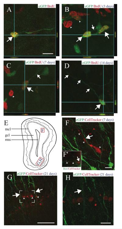
FIGURE 6. AORGs are born in the adult OB, and are closely apposed to migrating, immature neurons.
(A) Newborn AORGs (arrows) are found in the adult OB granule cell layer following adult BrdU administration. Co-labeling by BrdU (red) and eGFP (green) in AORGs was confirmed by three-dimensional confocal orthogonal reconstructions in the XZ and YZ planes. (B-D) Pulse labels of BrdU demonstrate that (B) AORGs (arrows) develop short processes (small arrow) by 3 days after their birth. Note eGFP-negative/BrdU-positive cells in (B) and (C) (arrowheads, putative adult-born granule neurons). (C) AORGs extend their processes further by 7 days (small arrow), and (D) display mature AORG morphology by 14 days after their birth. (E) Schematic representation of the olfactory bulb, indicating the regions from which panels F, G, and H are derived. (F) The whole cell label CellTracker Red was injected into the adult SVZa, revealing a close association between immature, adult-born migrating neurons (red, arrowheads) and the radial process of eGFP-positive AORGs (green, arrow) 7 days after CellTracker Red injection. (F’, inset) an example of a cell labeled by CellTracker Red that is immunopositive for doublecortin (Dcx, green), in the granule cell layer of the OB. Arrow indicates the cell body, and arrowheads mark the leading process of this migrating, immature neuron. (G and H) The close anatomical relationship between adult-born mature granule neurons (arrowheads) in the gcl and the radially oriented eGFP-positive fibers of AORGs (arrows) 21 days after CellTracker Red injection is shown via confocal reconstructions at lower (G; box indicates area magnified in (H)) and higher magnification (H). Scale bar in (A), 10 ⌈m for panels (A-D); scale bar in each of (F-H), 50 ⌈m. (gcl, granule cell layer; mcl, mitral cell layer; rms, rostral migratory stream). Schematic diagram based on Franklin and Paxinos (1997).

