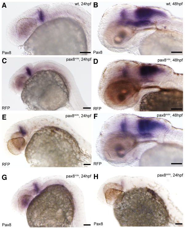
Figure 2.
Pax8 expression is identical to RFP expression. Transcripts were detected using in situ hybidization with either pax8 (A, B, G, H) or RFP (C, D, E, F) probes in 24hpf (A, C, E, G, H) or 48hpf (B, D, F) embryos. Expression pattern, at 24hpf, of pax8 in wild type (pax8+/+, A) was same to that of RFP in pax8+/m (C) and pax8m/m (E). Signal was detected in the mhb region of the embryos. RFP showed no signal in wild type samples (data not shown). At 48hpf the staining pattern of pax8 in wild type (B) resembled that of RFP expression in pax8+/m (D) and pax8m/m (F) embryos. Pax8+/m embryos showed positive staining also with the pax8 probe (G). Pax8 transcripts were also detected, at a much lower intensity, in pax8m/m embryos (H). Scale: 100 μm.

