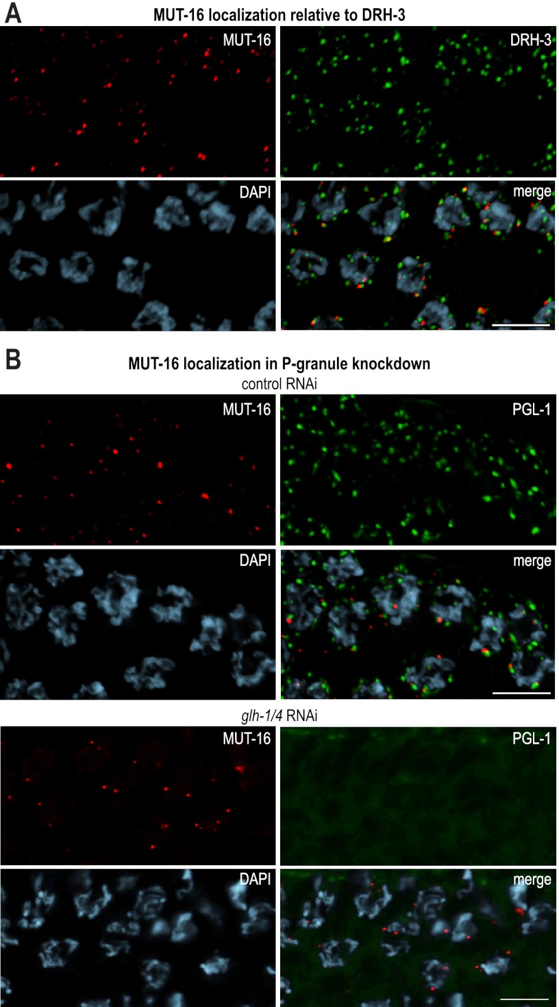
Figure 3.
Mutator proteins localize independently of P-granule components. (A) MUT-16 (red) and DRH-3 (green) form distinct foci adjacent to germline nuclei. Staining was performed using antibodies against GFP (MUT-16 in red), DRH-3 (green), and DAPI (blue). (B) MUT-16 and PGL-1 foci partially overlap in adult C. elegans feeding on Escherichia coli expressing control (empty vector) dsRNA. Upon treatment with glh-1/glh-4 dsRNA, PGL-1 becomes diffuse, but MUT-16 is unchanged. Proteins were visualized using anti-GFP (MUT-16 in red) and anti-dsRed (PGL-1 in green). DNA was stained by DAPI (blue). Bars, 5 μm.

