
Figure 2.
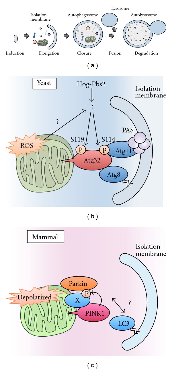
Mitophagy in yeast and mammals. (a) In autophagy, a double-membrane compartment, termed the isolation membrane, engulfs a portion of cytoplasm, including macromolecules and organelles, and forms autophagosomes. The late autophagosome fuses with the lysosome to form autolysosomes, which degrades the engulfed contents. (b) A model of mitophagy in yeast. Hog1 and Pbs2 regulate mitophagy by promoting the phosphorylation of Ser114 and Ser119 on Atg32. When Ser114 is phosphorylated, Atg32 binds to Atg11. Atg11 is a core component that facilitates the recruitment of specific cargoes to PAS, where the autophagosomes are generated. Atg32 also binds to Atg8, a factor that is essential for autophagosome formation. Autophagosomes envelop the mitochondria and fuse with lysosomes to degrade the mitochondria. (c) A model of mitophagy in mammalian cells. When mitochondria are depolarized, PINK1 accumulates on the mitochondrial outer membrane and recruits Parkin from the cytosol to the depolarized mitochondria in a manner dependent on the kinase activity of PINK1. The PINK1/Parkin complex triggers the autophagosome formation and the degradation of damaged mitochondria. Recent findings suggest that the proteasome is also involved in the destruction of mitochondria (see text for details).