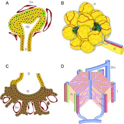
Fig. 1.
Close association of vessels with pancreas and liver epithelium throughout development into adulthood. (A) The early pancreatic bud is encased in capillaries (red) that pervade the gut mesenchyme. Endocrine cells (green) emerge within the bud epithelium (yellow). (B) The developing pancreatic tree grows coordinately with its vasculature, with vessels (red/blue) running along ducts. Islets (green) are embedded within the more abundant exocrine tissue (yellow). (C) The early liver bud shows intercalation of early hepatocytes (brown) and capillaries (red) within the septum transversum. (D) The developing liver contains a dense vascular network – the sinusoidal endothelium (purple). a, acini; bv, blood vessel; d, pancreatic duct; dp, dorsal pancreas; g, gut tube; h, hepatocytes; lb, liver bud; t, triad of hepatic artery, portal vein and bile duct; thv, terminal hepatic vein.

