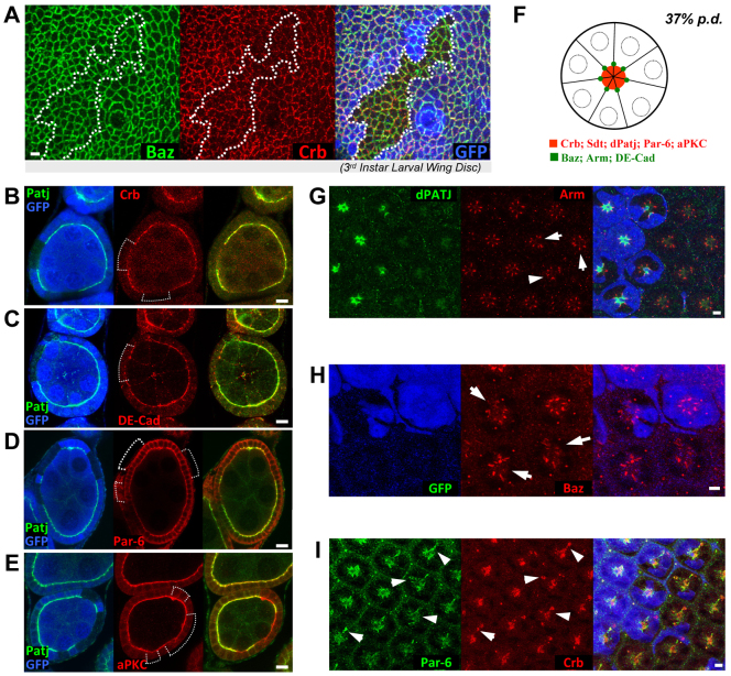
Fig. 3.
dPatj supports Crb expression in polarized epithelial cells and apical-basal polarity in early pupal photoreceptors. (A) dPatjΔ7 clone (outlined) in a larval imaginal disc shows subtle reduction in Crb expression but normal expression of Baz. (B-E) dPatjΔ7 clones in follicular epithelia show more marked loss of Crb (B) but no obvious disruption of DE-Cad (C), Par-6 (D) or aPKC (E). (F) Illustration of a transverse view of an early pupal ommatidium. Before 40% pupal development (pd), the apical membranes of all photoreceptors are converged at the center of the ommatidium and show enriched localization of Crb, Sdt, dPatj, Par-6 and aPKC (red). Adherens junctions that form between photoreceptors show ring-like patterns that are marked by DE-Cad, Arm and Baz (green). (G-I) In dPatjΔ7 mutant photoreceptors, Arm (G) and Baz (H) show mild disruptions (examples indicated by arrows), whereas Crb and Par-6 exhibit more severe mislocalization (I, arrowheads). All samples are from 37% pd pupae. In all panels, dPatjΔ7clones are marked by the loss of GFP (blue). Scale bars: 10 μm.

