Fig. 6.
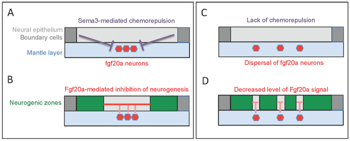
Model of the role of boundaries in neuronal positioning. A single zebrafish hindbrain segment is represented with the neural epithelium in light grey, boundary cells in dark grey, and the mantle layer in blue. (A,B) Normal embryos, in which Sema3fb/Sema3gb-mediated chemorepulsion from boundary cells positions fgf20a neurons (red) in the segment centre. The cluster of fgf20a neurons creates a focussed source of FGF signals (red lines) that inhibit neurogenesis in the adjacent neural epithelium, restricting the neurogenic zones (green) to being adjacent to boundaries. (C,D) Embryos in which boundary cells have been depleted or Sema3fb/Sema3gb signalling disrupted. The consequent dispersal of fgf20a neurons leads to disorganisation of the non-neurogenic zones and diminishes the level of FGF signalling.

