
Fig. 10.
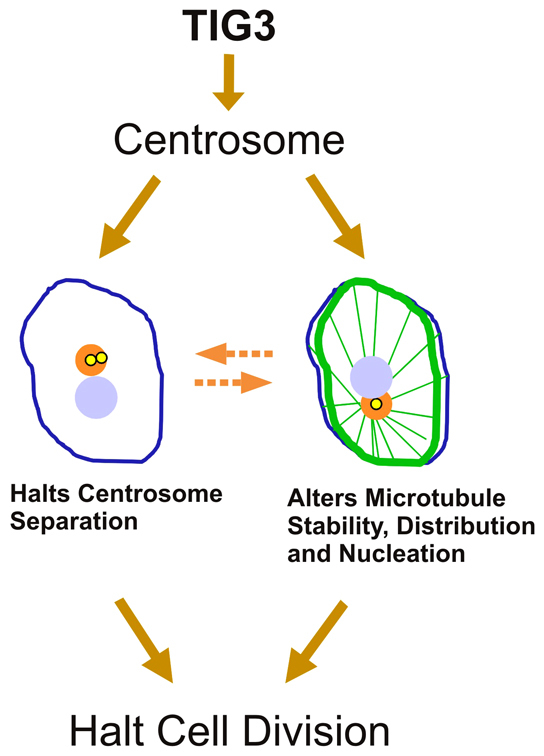
Schematic of TIG3 action. TIG3 interacts with the centrosome and halts centrosome separation during cell division. It also alters microtubule subcellular distribution, nucleation and stability. These changes result in the cessation of cell division. Centrosomes (yellow), nuclei (blue), TIG3 localization (orange) and microtubules (green) are shown. The blue outline represents the plasma membrane. The dashed arrows indicate that the centrosome and microtubules reciprocally influence the function of the other.