
Fig. 2.
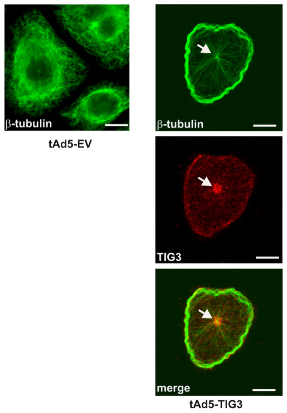
Impact of TIG3 on microtubule distribution. Keratinocytes were infected with an empty vector or TIG3-encoding virus, and after 24 hours fixed and stained with anti-β-tubulin (green) and anti-TIG3 (red) antibodies. The presence of TIG3 results in accumulation of β-tubulin in a band at the cell periphery (green). The arrow indicates accumulation of TIG3 at the centrosome.