
Fig. 5.
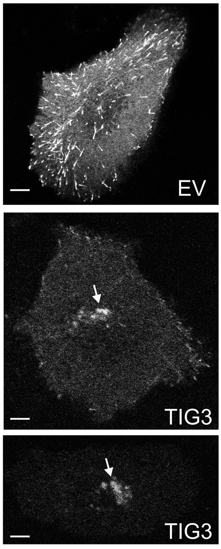
TIG3 reduces anterograde microtubule growth. Normal keratinocytes growing in glass-bottom dishes were transfected with 1 μg of EB1–GFP encoding plasmid in the presence of 2 μg of pcDNA3 (empty vector, EV) or pcDNA3-TIG3. After 18 hours, EB1–GFP fluorescence was detected using an Olympus FluoView FV1000 laser confocal microscope. The arrows indicate pericentrosomal distribution of EB1–GFP-labeled microtubules in two representative TIG3-positive cells. Scale bars: 10 μm.