
Fig. 7.
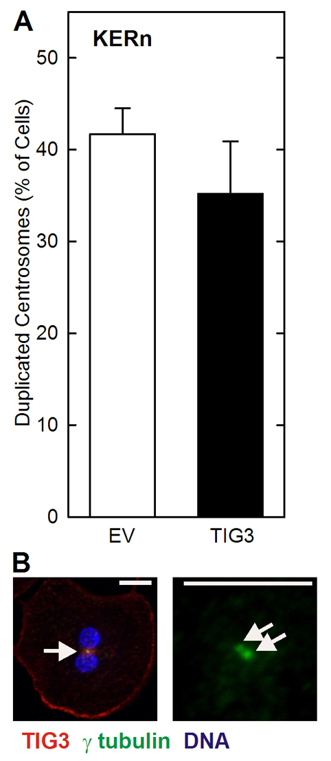
TIG3 does not inhibit centrosome duplication. (A) Centrosomes were counted in 30 EV or TIG3-expressing cells. A slight decrease in cells displaying duplicated centrosomes is observed in TIG3-expressing cells, but the reduction was not statistically significant (P<0.41). (B) Images showing centrosome duplication in a TIG3-expressing cell. Keratinocytes were infected with TIG3-expressing virus, and after 48 hours the cell was fixed and stained to detect TIG3 (red), γ-tubulin (green) and DNA (blue). The yellow staining in the left panel (arrow) indicates centrosome-localized TIG3. The arrows in the right panel show the closely spaced duplicated centrosomes in the same cell (only γ-tubulin staining is shown for clarity). Scale bars: 10 μm.