Fig. 8.
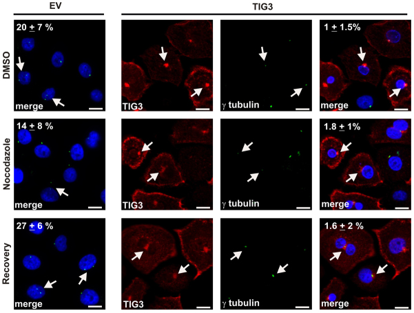
Keratinocytes were infected with tAd5-EV or tAd5-TIG3. After 24 hours, dishes of cells were treated with vehicle (untreated), 10 nM nocodazole for 2 hours (nocodazole), or nocodazole for 2 hours followed by 1 hour recovery in nocodazole-free medium (recovery). The cells were then fixed and stained with anti-γ-tubulin (green) and anti-TIG3 (red) antibodies, and stained with Hoechst 33258. The images were captured using a confocal microscope. Scale bars, 10 μm. The arrows indicate the centrosome. The counts are the number of cells that display a separated centrosome, and are presented as the mean ± s.d. derived from the count of ten separate fields.

