Fig. 9.
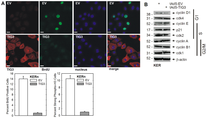
TIG3 suppresses cell division. (A) TIG3 reduces the incorporation of BrdU. Normal human foreskin keratinocytes were infected with empty or TIG3 encoding adenovirus, and after 24 hours they were incubated for 4 hours with BrdU. Cells were then fixed and stained with anti-TIG3 (red), anti-BrdU (green) antibodies and Hoechst 33258. The plots indicate the percentage of BrdU-positive cells (± s.e.m., n= four experiments with 50 cells counted per experiment) and the percentage of cells that stain strongly for phosphorylated histone H3 (± s.e.m. with 100 cells counted per experiment). Differences are significant as determined by Student's t-test, P<0.01. Scale bars: 10 μm. (B) Cell lysate was collected from keratinocytes at 24 hours after infection with tAd5-EV or tAd5-TIG3. The cell cycle regulatory-protein level was determined by immunoblot analysis. The numbers indicate the molecular weight (kDa). The phases of the cell cycle are indicated (G1, S, G2/M).

