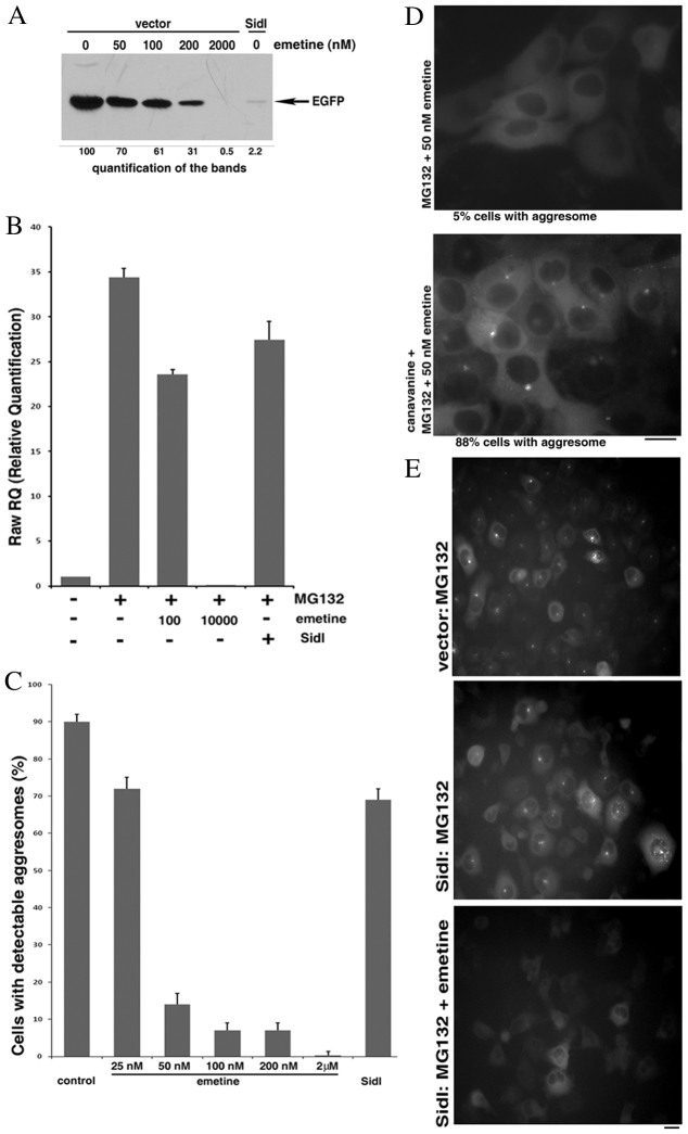
Fig. 4.
SidI reduces the threshold of DRiPs necessary to trigger aggresome formation. (A) Effects of various emetine concentrations and SidI on protein synthesis. HeLa cells were transfected for 3 hours with a plasmid encoding EGFP and co-transfected with either a plasmid encoding SidI or an empty vector. 1.5 hours after the end of the transfection indicated amounts of emetine were added to some samples. The levels of synthesized EGFP were assessed by immunoblotting 20 hours after the end of the transfection. (B) Divergent effects of SidI and emetine on induction of Hsp70. HeLa cells were transfected with a plasmid encoding SidI or an empty vector and on the next day incubated with or without 10 μM MG132 and the indicated concentrations of emetine for 7 hours. RNA was isolated from the cells and the levels of Hsp70 mRNA were assessed with RT-PCR. The effect of SidI was adjusted for the efficiency of transfection. (C) Divergent effects of SidI and emetine on aggresome formation. HeLa cells stably expressing Syn–GFP were transfected with a plasmid encoding SidI or an empty vector. On the next day cells were incubated for 4 hours with 10 μM MG132 and to some samples the indicated amounts of emetine were added; the aggresome formation was analyzed with a fluorescence microscope. The effect of SidI was adjusted for the efficiency of transfection. (D) Canavanine relieves inhibition of the aggresome formation by low concentration of emetine. Cells stably expressing Syn–GFP were incubated for 2 hours with 5 μM MG132 and 50 nM emetine with or without 20 mM canavanine. The extent of the aggresome formation was analyzed with a fluorescence microscope. (E) The remaining protein synthesis is necessary for aggresome formation in the presence of SidI. HeLa cells stably expressing Syn–GFP were transfected with a plasmid encoding either SidI or an empty vector. On the next day, cells were incubated for 4 hours with 10 μM MG132 and 5 μM emetine was added to one sample. The extent of the aggresome formation was analyzed with a fluorescence microscope. Scale bars: 20 μm.

