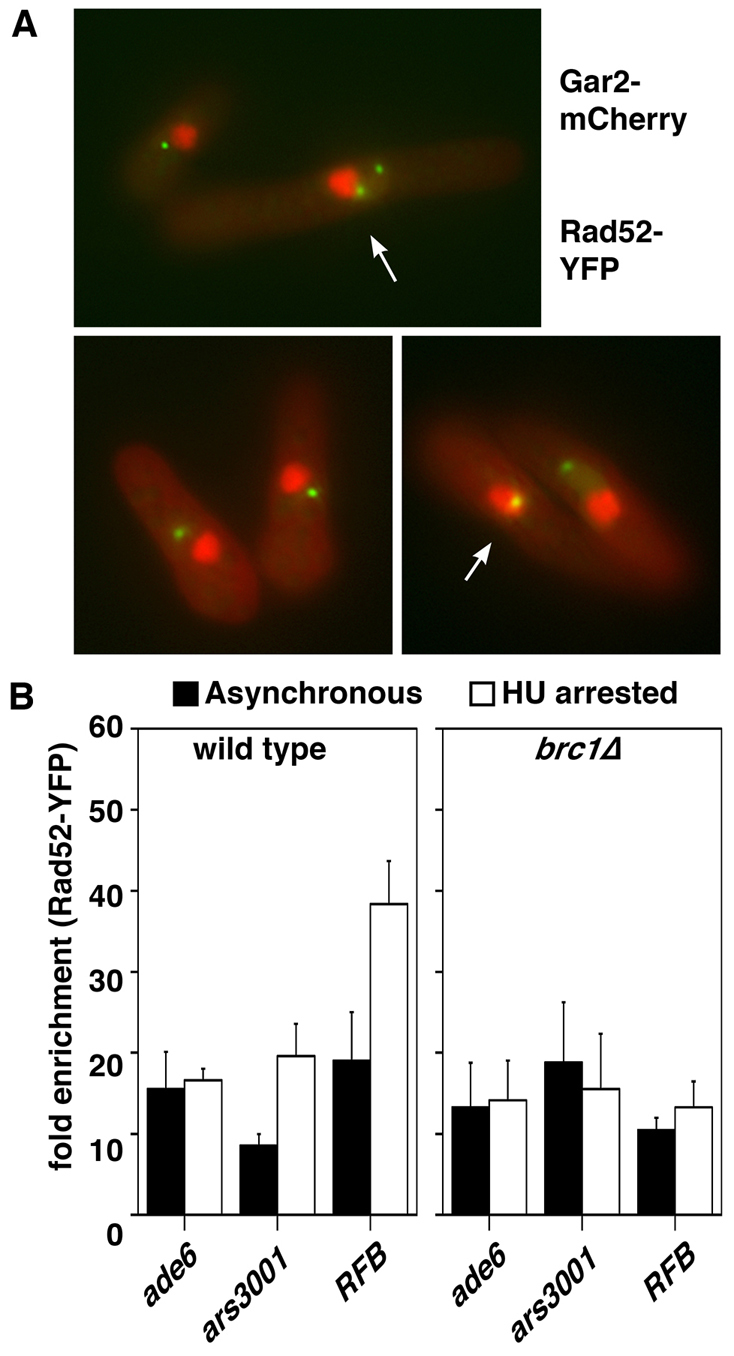
Fig. 7.

brc1Δ cells do accumulate Rad52 in the rDNA. (A) Rad52–YFP and Gar2–mCherry were simultaneously imaged in brc1Δ cells by conventional fluorescence microscopy. Colocalizing signals (arrowed) were observed in 15% of cells (n=50). (B) Anti-GFP ChIP analysis of the indicated loci from wild-type and brc1Δ cells in a Rad52–YFP background from either asynchronous cultures (solid bars) or following arrest in 11 mM HU (open bars) for 4 hours. Increased rDNA enrichment is evident in wild-type, but not in brc1Δ cells. Data are means ± s.e.m., n=3.
