
Figure 2.
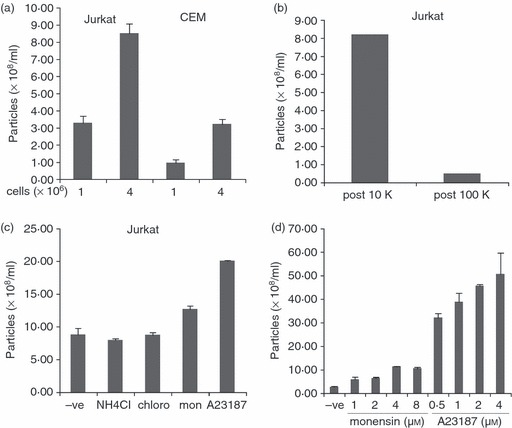
Quantitative determination of microvesicle release by nanoparticle tracking analysis (NTA). (a) 1 × 10⁶ and 4 × 10⁶ Jurkat and CEM cells were incubated in serum-free medium overnight. Post-10 000 g supernatant was analysed by NTA. The mean size of particles detected was 161 nm. (b) Conditioned medium from Jurkat cells incubated overnight in serum-free medium was analysed by NTA after 10 000 g and 100 000 g centrifugation. (c) Jurkat cells were incubated overnight in serum-free medium containing 50 mm NH4Cl, 100 μm chloroquine, 4 μm monensin, or 0·5 μm A23187. Post-10 000 g supernatant was analysed by NTA. The mean size of particles detected was 114 nm. (d) Jurkat cells were incubated overnight with the indicated concentrations of monensin and A23187. Post-10 000 g supernatants were analysed by NTA. The mean size of particles detected was 126 nm.