
Figure 3.
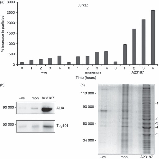
Ionophore treatment of Jurkat cells releases microvesicles containing exosome-associated polypeptides. Jurkat cells were treated for the indicated times with 4 μm monensin or 1 μm A23187. Post-10 000 g supernatants were analysed by nanoparticle tracking analysis (NTA). (b) Supernatants of Jurkat cells treated with the same concentration of ionophores as in (a) for 6 hr were spun at 10 000 g and then 100 000 g, and the latter pellets were analysed by immunoblotting for Alix and Tsg101 expression. The mean size of particles detected was 158 nm (c). Supernatants of Jurkat cells incubated for 6 hr as in (b) were spun at 10 000 g and 100 000 g and the latter pellets were analysed using SDS–PAGE and SimplyBlue protein staining. The indicated bands were excised and mass spectrometric fingerprinting was performed. Proteins identified included 1, heat-shock protein 90; 2, tubulin; 3, elongation factor α1; 4, actin; and 5, glyceraldehyde 3-phosphate dehydrogenase.