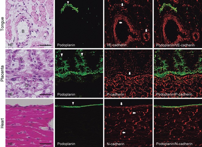
Fig. 1.

Immunostaining for podoplanin, P-, N- and VE-cadherin in mouse tissue. The immunostained sections were re-stained by hematoxylin-eosin (H-E) staining, which shows blood vessels (B) of the muscle layer of the tongue, the villus (V), and decidua (D) of the placenta, and the cardiac muscle and epicardium of the heart. The fluorescence with anti-podoplanin (arrowhead) is found on the lymphatic vessel of the tongue, on syncytiotrophoblasts of the placenta villus, and on the epicardium. The fluorescence with anti-VE-cadherin (arrows) is found on blood vessels, and weakly on the lymphatic vessel of the tongue. The lymphatic vessel immunostained with both anti-podoplanin and anti-VE-cadherin is shown in the merged image. The fluorescence with anti-P-cadherin (arrows) is found on the decidua of the placenta. The separation of the area immunostained with anti-podoplanin and anti-P-cadherin in the placenta is shown in the merged image. The fluorescence with anti-N-cadherin (arrows) is found on epicardium and intercalated disks without a cross-reaction with cardiac muscle. The epicardium immunostained with both anti-podoplanin and anti-N-cadherin is shown in the merged image. The anti-podoplanin and ant-cadherins did not show a cross-reaction with connective tissue or muscle. Bar: 50 μm.
