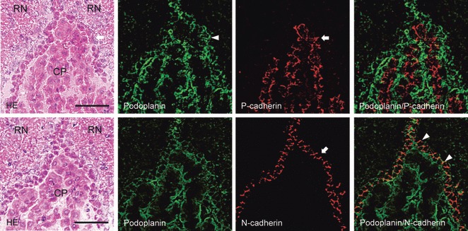
Fig. 3.

Immunostaining for podoplanin, P- and N-cadherin of the third ventricle. The double immunostained section for podoplanin and P-cadherin is adjacent to the double immunostained section for podoplanin and N-cadherin. Immunostained sections were re-stained by H-E staining, which shows red nuclei (RN), choroid plexus (CP), and ependymal cells (arrow). The fluorescence with anti-podoplanin is found on ependymal cells (arrow) and choroid plexus epithelia. The fluorescence with anti-P-cadherin (arrowhead) is found inside the choroid plexus, where anti-podoplanin did not react in the merged image. The fluorescence with anti-N-cadherin is also found on ependymal cells (arrow) where anti-podoplanin reacted (arrowheads) but was absent on choroid plexus epithelia in the merged image. Bar: 50 μm.
