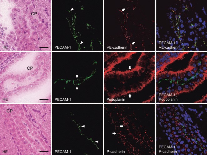
Fig. 6.

The laser-scanning confocal microscopy of immunostaining for podoplanin, P- and VE-cadherins on the choroid plexus fibrovascular core of the lateral ventricle. The upper, middle, and lower panels are the same sections. The immunostained sections were re-stained by the H-E staining, which shows the choroid plexus (CP). In the immunostaining with anti-PECAM-1 and anti-VE-cadherin, the fluorescence with anti-VE-cadherin is present on the choroid plexus endothelial cells (arrows) of the fibrovascular core as well as the region reacted with anti-PECAM-1 (arrowheads). The region reacting with anti-PECAM-1 coincided with the region reacting with anti-VE-cadherin in the merged image. In the immunostaining with anti-PECAM-1 and anti-podoplanin, there is fluorescence with anti-PECAM-1 on the choroid plexus endothelial cells (arrowheads) of the fibrovascular core, whereas fluorescence with anti-podoplanin occurs on the choroid plexus epithelial cells (arrows) on the ventricle side both on the cell surface and in the cell–cell junctions. The region reacting with anti-PECAM-1 did not coincide with the region reacting with anti-podoplanin in the merged image. In the immunostaining with anti-PECAM-1 and anti-P-cadherin, the fluorescence with anti-P-cadherin is on the choroid plexus epithelial cells (arrows) at the basement membrane side both on the cell surface and in the cell–cell junctions, whereas the fluorescence with anti-PECAM-1 is on the choroid plexus endothelial cells (arrowheads) of the fibrovascular core. The region reacting with anti-PECAM-1 did not coincide with the region reacting with anti-P-cadherin in the merged image. Bar: 20 μm.
