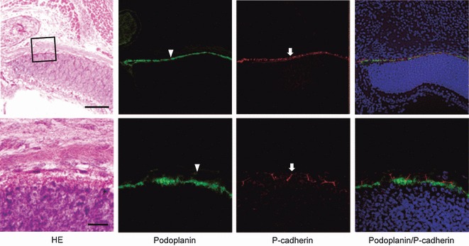
Fig. 7.

Immunostaining for podoplanin and P-cadherin of the retina. Upper sections were examined by a fluorescence microscopy (bar: 100 μm) and lower sections by laser-scanning confocal microscopy (bar: 20 μm). The fluorescence with anti-podoplanin is found on the surface of retinal pigment epithelial cells (arrowheads), whereas the fluorescence with anti-P-cadherin is localized on retinal pigment epithelial cells at cell–cell junctions (arrows).
