Fig. 8.

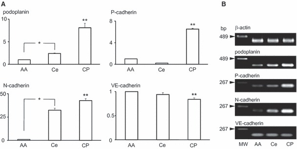
Gene analysis for the expression of podoplanin, P-, N-, and VE-cadherin mRNAs in the choroid plexus. Total RNA extraction from tissue of the abdominal aorta (AA), the surface of cerebrum with the pia mater (Ce), and the ventricular wall with choroid plexus (CP) were examined. (A) Real-time PCR analysis. Relative mRNA amounts are expressed in arbitrary units. Target gene cDNA units in each sample were normalized to β-actin cDNA units. Podoplanin, P- and N-cadherin mRNA amounts were significantly larger in the ventricular wall with choroid plexus than in cerebrum tissue or in abdominal aorta as a negative control. The VE-cadherin mRNA amount was significantly smaller in the ventricular wall with choroid plexus than in cerebrum tissue and in abdominal aorta as a positive control. The amounts of podoplanin and N-cadherin mRNA were significantly larger in the surface of cerebrum with the pia mater than in abdominal aorta. *Statistically significant difference by Student’s t-test and one-way anova** (P < 0.01). (B) RT-PCR analysis. Intensities of amplicon for β-actin mRNA and VE-cadherin were similar in abdominal aorta, cerebrum, and ventricular wall with choroid plexus. Intensities of amplicon for podoplanin, P- and N-cadherin mRNA increased in strength in the order ventricular wall with choroid plexus, cerebrum, and abdominal aorta.
