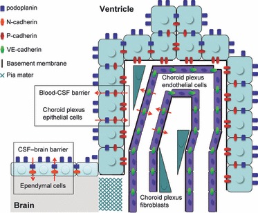
Fig. 9.

A schematic outline of the expression for podoplanin, P-, N-, and VE-cadherin in the choroid plexus. Podoplanin is expressed on the cell surface and at the cell–cell junctions of ependymal cells and choroid plexus epithelial cells. P-cadherin is expressed on choroid plexus epithelial cells at the vascular side between epithelial cells and endothelial cells or fibroblasts. VE-cadherin is expressed on choroid plexus endothelial cells at the cell–cell junctions. N-cadherin is expressed on the pia mater and ependymal cells at the cell surface and cell–cell junctions. Podoplanin and P-cadherin participate in the blood-CSF barrier, and podoplanin and N-cadherin play roles in the CSF-brain barrier.
