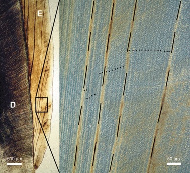
Fig. 2.

Transmitted light microscope images of incremental markings in buccal enamel (E) of a goat M2. Higher magnification image (right frame) viewed with phase contrast. Accentuated incremental lines (Wilson bands, dashed lines) that were used as landmark lines exhibit a variable spacing. Regular (daily) incremental lines (laminations) are indicated by dots. D, dentin. Cuspal direction to top of images.
