
Fig. 9.
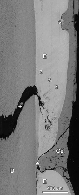
Aplastic defect (lower asterisk) and hypoplastic defect (upper asterisk) in lingual enamel (E) of a goat M2. Both defects are occluded by cementum (Ce) and were not correctly diagnosed on macroscopic inspection of the tooth surface. D, dentin; +, sectioning artifact. BSE image. Numbering of Wilson bands corresponds to that in Figs 6 and 7. Cuspal direction to top of image.