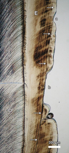
Fig. 10.

Transmitted light microscope image showing hypoplastic defects (a–c) in the buccal enamel (E) of an M1 of a sheep or goat (species assignment questionable). All defects are associated with the same accentuated incremental line (Wilson band, arrows) and were thus formed contemporaneously. D, dentin. Cuspal direction to top of image.
