
Fig. 1.
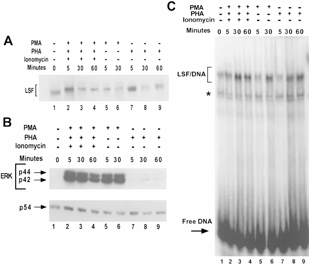
Mitogenic stimulation of human peripheral T cells leads to LSF phosphorylation, increased LSF-DNA binding activity, and ERK activation. A: Western blot analysis of nuclear extracts from either unstimulated or stimulated human peripheral T cells. Approximately 40 µg of extract were loaded per lane. The blot was probed with α-LSF pep1-1 antibody. Lane 1: unstimulated cells, lanes 2–4: cells treated with PHA, ionomycin, and 74 ng/ml of PMA, lanes 5–6: cells treated with 74 ng/ml of PMA, and lanes 7–9: cells treated with PHA. The lengths of time of treatment in min are indicated. Note the slight curvature across the lanes in the relative mobilities of the proteins. B: Kinetics of ERK activation by mitogenic stimuli in T cells. Twenty micrograms of the same samples used in panel A were loaded per lane. The blot was probed with antiphospho-ERK antibody (upper panel) and reprobed with JNK antibody (lower panel) to detect differences in the loading of total protein. Lanes 1–9 are as in panel A. The positions of phosphorylated p44 ERK1, phosphorylated p42 ERK2, and p54 JNK are indicated. Note the slight curvature across the lanes in the relative mobilities of the proteins. C: LSF DNA-binding activity is enhanced in response to mitogenic stimulation in T cells. EMSA of nuclear extracts prepared in a separate experiment from that in panels A and B, either from quiescent cells or from cells stimulated with various mitogens. The positions of migration of LSF/DNA complexes and free DNA are indicated. The asterisk denotes a complex with non-specific DNA-binding proteins. In this experiment, increases in LSF DNA-binding activity were 2.3-fold (lane 3), 1.9-fold (lane 6), and 1.4-fold (lane 9), as compared with lane 1.