
Fig. 4.
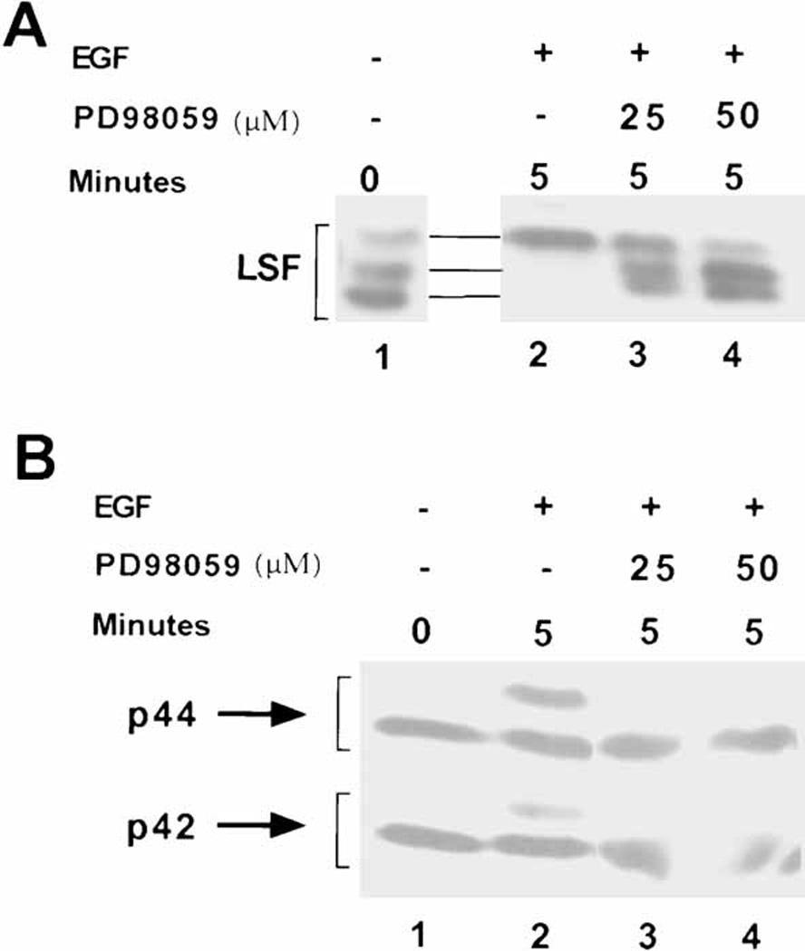
The ERK pathway targets LSF for phosphorylation after mitogenic stimulation of NIH 3T3 cells. A: Western blot analysis of LSF, as described in Fig. 2. Lane 1: extract from serum-deprived NIH 3T3 cells; lanes 2–4: extracts from NIH 3T3 cells stimulated with EGF for 5 min. Lanes 3,4: cells were pre-treated with the MEK1/2 inhibitor PD98059, at the indicated concentrations. Note the slight curvature across the lanes in the relative mobilities of the proteins. B: Western blot analysis of the samples in panel A with polyclonal antibody against ERK1. This antibody recognizes both inactive and active (slower migrating) forms of both ERK1 and ERK2.