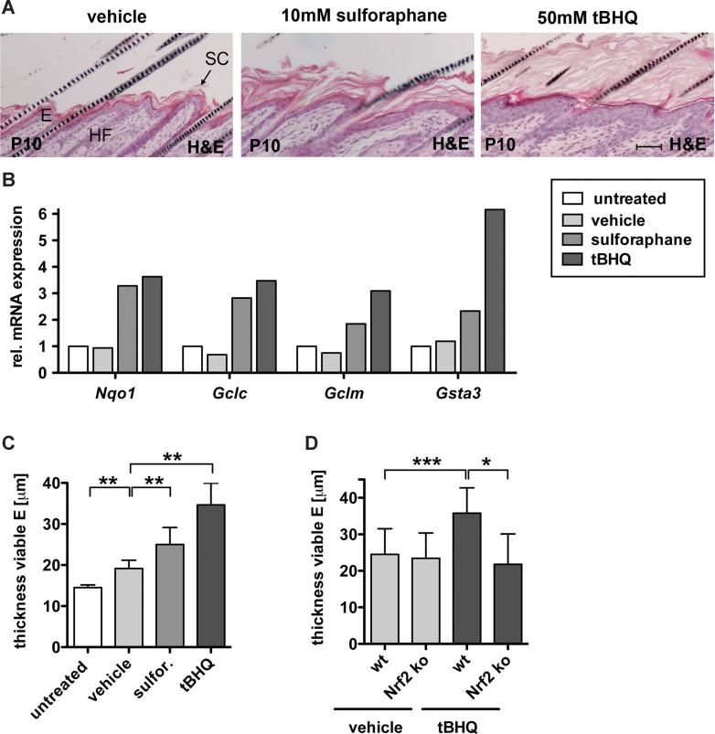
H&E staining of longitudinal skin sections from mice topically treated with vehicle, 10 mM sulforaphane (sulfor) or 50 mM tBHQ showing hyperkeratosis and acanthosis in sulforaphane and tBHQ treated mice. Scale bar, 50 µm. E, epidermis; HF, hair follicle; SC, stratum corneum.
qRT-PCR analysis of Nqo1, Gclc, Gclm and Gsta3 relative to Gapdh using RNAs from back skin of the differently treated mice (control, N = 3; vehicle, N = 3; sulforaphane, N = 3; tBHQ, N = 3). Expression levels in untreated mice were arbitrary set to 1 (dashed line).
Thickness of the viable epidermis in untreated (N = 10), vehicle (N = 6, **p = 0.0022), sulforaphane (N = 6, **p = 0.0022) or tBHQ (N = 6, **p = 0.0012) treated WT mice.
Thickness of the viable epidermis in WT and Nrf2 ko mice treated with vehicle or tBHQ. Epidermal thickness increased in WT mice (N = 15/17, ***p = 0.0003), but not in Nrf2 ko mice (N = 17/5, **p = 0.0118).

