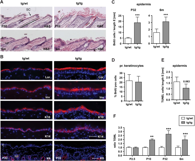
H&E staining of P32 (upper panel) and 6 months (lower panel) K5cre-CMVcaNrf2 and control skin sections. Scale bar, 100 µm.
Immunofluorescence analysis of Lor, Inv, K10, K14 and K6 (red), counterstained with Hoechst (blue). Scale bars, Lor, Inv, K10 and K14, 30 µm; K6, 50 µm. E, epidermis; HF, hair follicle; SC, stratum corneum.
BrdU positive cells per length epidermis at P32 (***p = 0.0004) and 6 months of age (***p = 0.0004).
Percentage of BrdU positive subconfluent primary keratinocytes from tg/wt and tg/tg mice.
TUNEL positive cells per length epidermis at P32 (N = 4/5, p = 0.063).
TEWL at P2.5 (N = 4/2), P10 (N = 8/4, **p = 0.0016), P32 (N = 10/6, ***p = 0.0002) and 6 months (N = 11/12, ***p < 0.0001).

