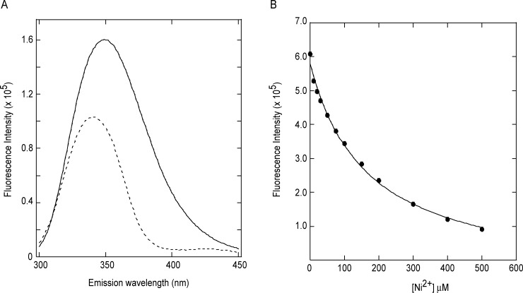
Figure 6.

Intrinsic tryptophan fluorescence data indicates direct binding of Ni2+ to gp74. (A) Emission spectra of 1 μM gp74 in the absence (solid line) and presence of 1M Ni2+ ions (dotted line). (B) Binding of Ni2+ ions to gp74 as monitored by intrinsic tryptophan fluorescence. The data (solid circles) were fit assuming a 1:1 complex (solid line),42 as described in Fluorescence metal-binding studies.
