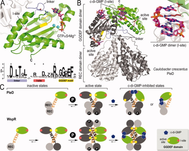
Figure 4.
GGDEF domains and diguanylate cyclases. (A) Prototypical GGDEF domain structure. The PleD GGDEF domain bound to GTP-alpha-S is shown (PDB code: 2V0N).44 The GGDEF motif is colored yellow. The position of the I-site is highlighted as small spheres. The sequence logo highlights several conserved motifs extending in to the linker upstream of the GGDEF domain fold. (B) Product-inhibited structure of full-length PleD. The structure of a PleD dimer activated by BeF−3 bound to one REC domain is shown. The enzyme is inhibited by a stacked c-di-GMP dimer that occupies the I-site on the GGDEF domains (inset). (C) Models for the regulatory cycle for PleD and WspR. The models highlight conserved and unique feature that control enzymatic function of the two proteins.

