
Figure 3.
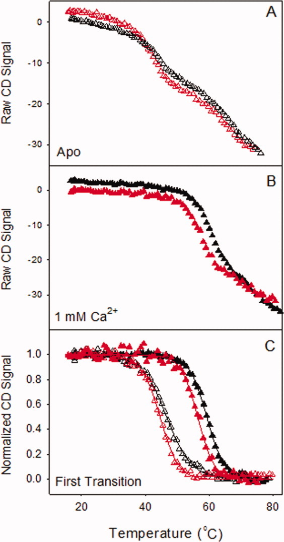
Thermal-unfolding of wild-type and X-enabled NCAD12 at 230 nm. The raw CD signal versus the probe temperature is represented for wild-type (black) and X-enabled NCAD12 (red) in (A) the apo-state (Δ) and (B) the calcium saturated state (▴). (C) Normalized data from the first transition for the apo and calcium-bound states are plotted versus temperature. The solid lines are simulated based on parameters resolved from fits to the Gibbs–Helmholtz equation.