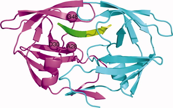
Figure 1.

The predicted sites of mutations in the specificity designed Pr3 (A28S, D30F, G48R) HIV-1 protease. In this single-chain HIV protease structure, the sequences corresponding to the two monomers are colored differently: monomer A is in magenta and monomer B is in cyan. The substrate is shown in green with respect to the scissile bond; N terminal residues colored green and C terminal residues colored limon green.
