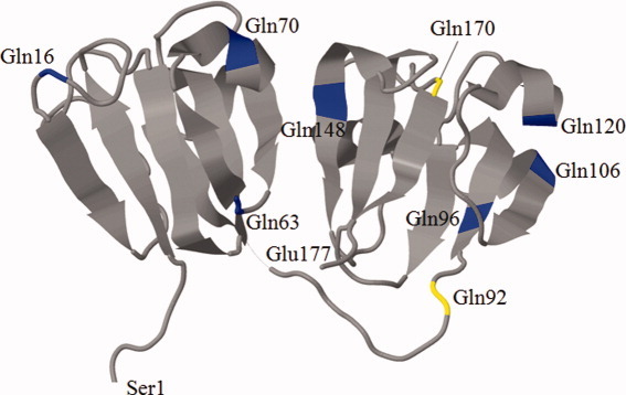
Figure 4.

A three-dimensional representative structure of murine γS crystallin obtained from the Protein Data Bank (PDB; http://www.rcsb.org/pdb/home/home.do) showing all possible Gln residues in γS crystallin (blue) with Gln residues 92 and 170 (yellow). [Color figure can be viewed in the online issue, which is available at wileyonlinelibrary.com.]
