
Figure 1.
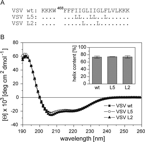
Secondary structure of VSV TMD peptides. A: Primary structures of peptides. Hydrophobic residues of the VSV G-protein (strain San Juan) TMD are flanked by Lys-triplets to enhance solubility and a Trp is added for quantification. Dots correspond to wild-type residues. B: CD spectra and calculated percentages of helix structure (inset); residual secondary structure is accounted for by β-sheet (5–6%), β-turn (∼12%), and random coil (8–9%). All values represent means of three independent measurements + SD.