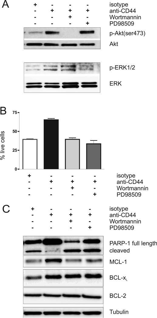
Figure 4. The anti-apoptotic effect of CD44 on CLL-cells is blocked by PI3K/AKT or MAPK/ERK inhibitors.
CLL cells were pre-incubated either with the PI3K inhibitor, wortmannin (100nM), or with the MEK inhibitor, PD98509 (50μM), for 30 minutes. Then cells were incubated with either an isotype control antibody (anti-mouse IgG2, 10μg/ml) or a CD44 activating antibody (BU75, 10μg/ml) for 30 minutes, washed and incubated with secondary anti-mouse antibody (1μg/ml). (A, C) Cell lysates (>90% CLL cells) were obtained at baseline and at 30 minutes after stimulation and were then subjected to Western blot analysis. (A) Activation the AKT and ERK. (B) Cell viability by MitoTracker staining. The mean and standard deviation of 3 independent experiments is shown (mean % live cells: isotype 39 ±1%, CD44 stimulated 65 ±3%, CD44 stimulated plus Wortmannin 40 ±3%, CD44 stimulated plus PD98509 34 ±8%, p<0.0.02 for comparisons of CD44 stimulated to control or drug treated cells, p>0.25 for comparisons of isotype treated to CD44 stimulated cells in the presence of inhibitors). (C) Expression of BCL-2 family members.

