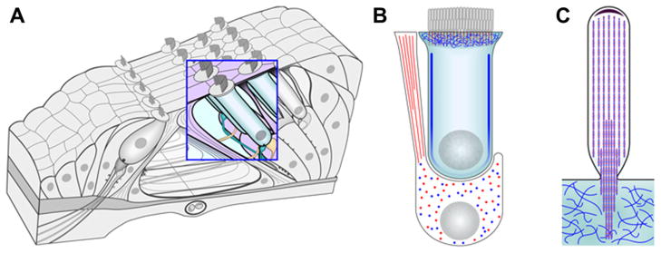
Fig. 2.
β-actin and γ-actin are differentially distributed in hair and supporting cells in the organ of Corti. The distribution of the two cytoplasmic actins in the organ of Corti (A) was determined by immuno transmission electron microscopy (TEM) (Hofer et al., 1997; Furness et al., 2005) and is summarized in this schematic. Red is used to signify β-actin and blue for γ-actin. An enlargement of an outer hair cell and Deiters’ cell (B) show that the phalanges of Deiters’ cells are primarily β-actin based, but the remainder of the cell body contains a relatively homogenous composition of the two cytoplasmic actins. In contrast, the lateral wall and cuticular plate of the hair cell is enriched for γ-actin. In adult guinea pig stereocilia (C) the total estimated ratio of γ-actin to β-actin is 2:1, though locally high concentrations of β-actin exist. The unidirectional paracrystalline array of F-actin in the stereocilium is illustrated as being composed of both γ-actin and β-actin, but the specific distribution of the two remains unclear and is represented here as a copolymer.

