
Figure 3.
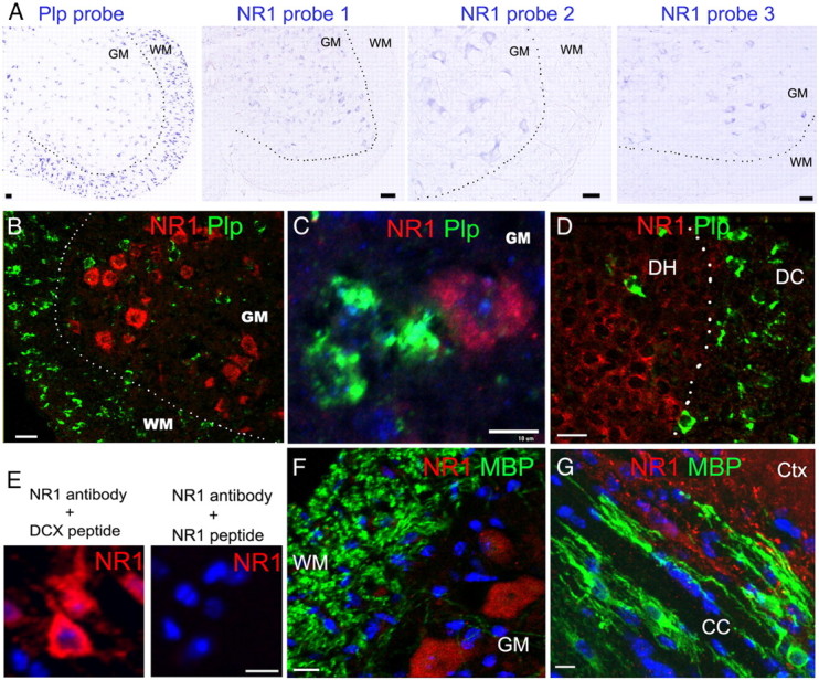
mRNA ISH and immunohistochemistry showing barely detectable NR1 transcript and protein in oligodendrocytes. A, Single mRNA ISH in P8∼P10 spinal cord, revealed by alkaline phosphatase-mediated color development, shows NR1 transcripts in GM, but barely detectable in WM (right three panels), despite presence of many Plp+ oligodendrocytes (left panel). B–D, Confocal images of dual fluorescent mRNA ISH demonstrating NR1 (pooled four NR1 probes) and Plp transcripts in P8∼P10 spinal WM and GM (B, C), and in dorsal column (DC) and dorsal horn (DH) (D). Note that Plp+ oligodendrocytes express barely detectable NR1 transcripts. E, NR1 peptide-antibody neutralization experiments demonstrating the specificity of NR1 immunostaining signals in cortical neurons. DCX, Doublecortin. F, G, NR1 and MBP double immunohistochemistry showing barely detectable NR1 signals in MBP+ myelin and oligodendrocytes from PRG mice treated with vehicle. Note the strongly positive NR1 signals in spinal GM (F) and forebrain cortex (Ctx) (G). CC, Corpus callosum. Scale bars, 10 μm, applied to all.