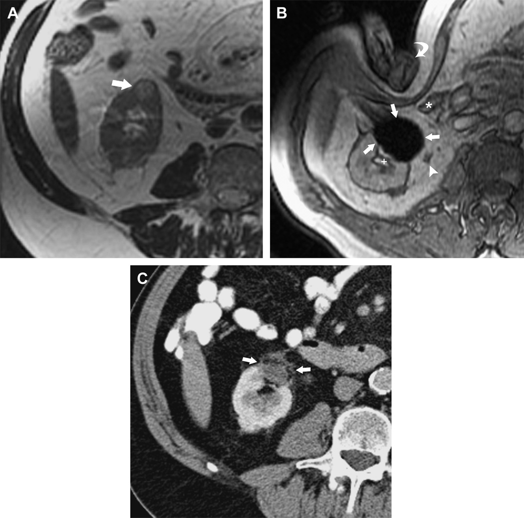
Figure 11.
MRI-guided Percutaneous Cryotherapy of Renal Tumors
70-year-old man with renal cell carcinoma of the right kidney lower pole treated with MRI-guided percutaneous cryoablation. (a) Transverse T2-weighted FRFSE image obtained before treatment in 1.5 T MRI shows a small exophytic renal mass in the lower pole of the right kidney anteriorly (arrow). (b) Intraprocedural transverse gradient echo image obtained in 0.5 T open configuration interventional MRI shows that sharp edge definition of signal void ice ball (arrows) contributes to monitoring of tumor coverage and assessment of proximity to adjacent ureter (arrowhead), renal collecting system (+), and colon (*) which is being displaced by the interventionalist's hand (curved arrow). (c) 18 month follow-up contrast enhanced transverse CT image shows no enhancement in the involuted ablation zone (arrows).

