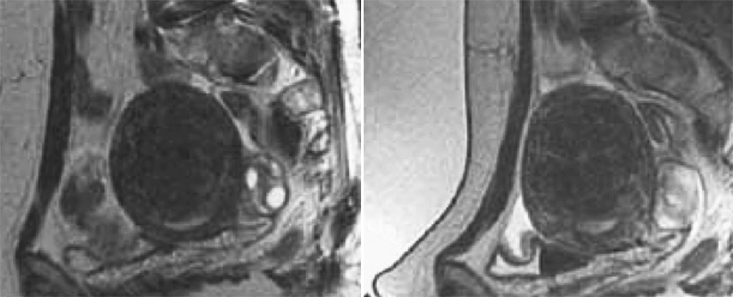
Figure 2.
The sagittal localizer image on the left demonstrates bowel loops coursing between the anterior abdominal wall and the uterine fibroid. After placement of a spacer device (sagittal localizer image on the right) under the anterior abdominal wall, the bowel loops are displaced, allowing for treatment through a larger acoustic window. (Reproduced, with permission, from Lippincott Williams and Wilkins. “A review of magnetic resonance imaging-guided focused ultrasound surgery of uterine fibroids", Top Magn Reson Imaging. 2006 Jun;17(3):173–9).

