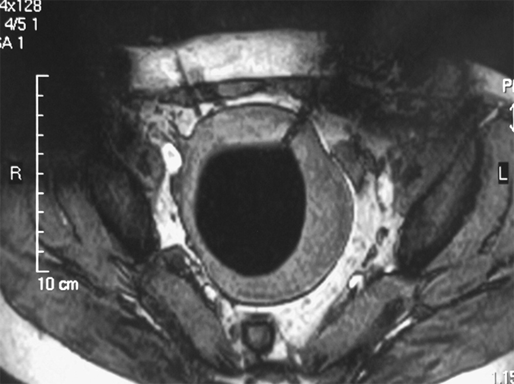
Figure 5.
Axial T2 weighted spin echo sequence demonstrating a probe in the left anterolateral aspect of a uterine fibroid. The diffuse low signal intensity in the fibroid represents the ice-ball. (Image courtesy of Yusuke Sakuhara, MD, Department of Radiology, Hokkaido University Hospital, Sapporo, Japan)

