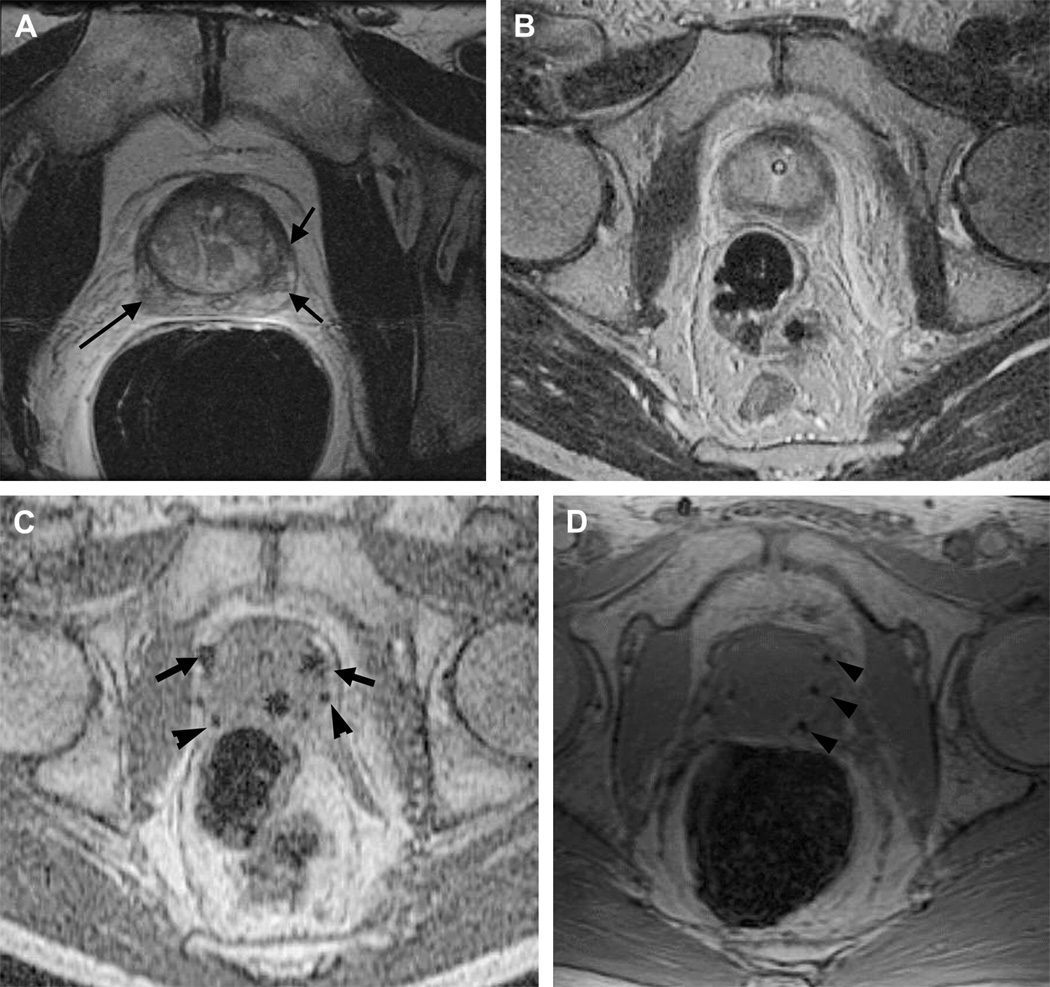
Figure 7.
Pre-, intra-, and post-operative MRI-guided brachytherapy in prostate cancer.
Pre-operative 1.5T (A) axial T2 weighted spin echo image through the prostate base, demonstrating low signal intensity in the peripheral zone (arrows), previously demonstrated to be tumor. Intra-operative 0.5T (B) axial T2 weighted spin echo T2 weighted spin echo image through the same area. Intra-operative axial gradient echo MR images (C) obtained in real-time during needle and seed placement in the prostate base. The larger round areas represent the needles (arrows), prior to deployment, and the small round areas represent the deployed seeds (arrowheads). A post-operative axial SPGR through the prostate base demonstrates multiple round areas of low signal in the peripheral zone (arrowheads), consistent with deployed seeds.

