
Figure 6.
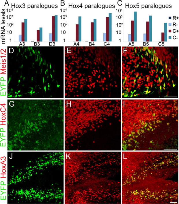
Verification of Hox gene expression in caudal 5HT neurons. A–C, Taqman RT-PCR confirmed the expression of Hox3 (A), Hox4 (B), and Hox5 (C) paralogues in caudal (C+) but not rostral (R+) 5HT neurons. D–L, Immunohistochemical staining for the Hox cofactors, Meis 1 and Meis 2, and Hox proteins. D–F, Meis1 and Meis2 (E) were detected in caudal 5HT neurons (D) with a pan-anti-Meis antibody (F, overlay). G–I, HoxC4 protein (H) was detected in caudal 5HT neurons (G, I, overlay). J–L, HoxA3 protein (K) was detected in most caudal 5HT neurons (J) at E12.5 (L, overlay). Scale bars: D–L, 50 μm.