
Fig. 1.
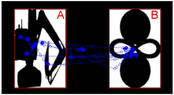
Visual examination behavior in the VPC test phase. In this representative example, the familiar image is on the left (A), and the novel image is on the right (B), for a normal control subject. The detected gaze positions are indicated by blue circles, with the connecting lines indicating the ordering of the gaze positions.