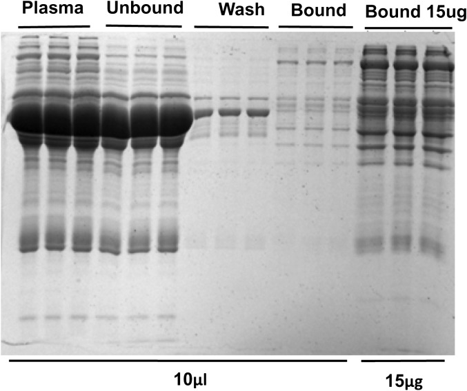
Figure 1.
After plasma was treated with concanavalin A (ConA) lectin, sodium dodecyl sulfate–polyacrylamide 1-dimensional gel electrophoresis was performed. The gel was loaded with 0.5 μL of untreated plasma diluted in ConA buffer, the unbound fraction, the wash, which was retained for this analysis, and the bound fractions. Lanes were loaded in equivolume amounts (10 μL) or on the basis of equal protein (15 μg per lane), as labeled. Gels were then stained with Coomassie blue.

