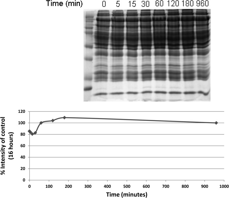
Figure 2.
Sodium dodecyl sulfate (SDS)–polyacrylamide 1-dimensional gel electrophoresis performed after incubating obtained after plasma was allowed to incubate on concanavalin A lectin columns for varying amount of times, in minutes, as shown. After separating an equal volume of bound fraction by SDS gel electrophoresis, the total amount of protein in each lane was determined by calculating the lane intensity after staining with Coomassie blue and plotted. The percent intensity of the control condition (overnight incubation, or 16 hours) is plotted for each time condition.

