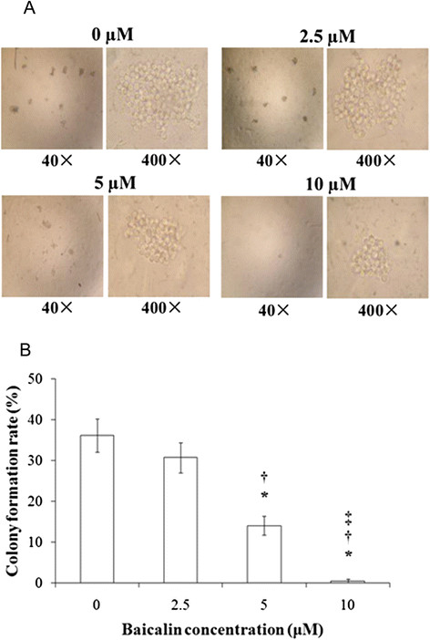
Figure 2.
Formation of CA46 cell colonies after treatment with baicalin at varying concentrations. Cells (4 × 10²/well) were cultured with baicalin at the indicated concentrations for 10 days. Colony formation rates were determined as described in Materials and methods. Sampling was performed in triplicate for each experimental condition. (A), phase contrast inverse microscopy. (B), colony formation rates with findings presented as means ± standard deviation for three independent experiments. *P <0.01 compared to the solvent control; †P <0.01 compared to 2.5 μM baicalin; ‡P <0.01 compared to 5 μM baicalin.

