Figure 3.
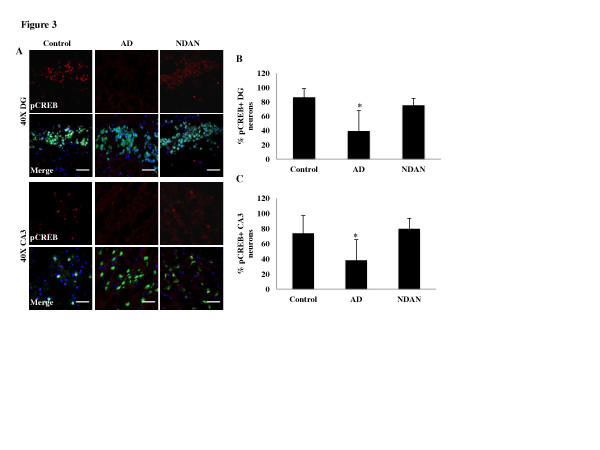
Synaptic integrity is maintained in the NDAN hippocampus as suggested by preserved levels of neuronal nuclear pCREB. (A) pCREB (red) and NeuN (green) expression was assessed in 10 μm sections of hippocampus. DAPI-containing mounting medium was used to visualize nuclei (blue). pCREB immunoreactivity was decreased in the DG and CA3 neurons of AD hippocampi but not in NDAN cases (scale bar 50 μm). The number of neurons positive for nuclear pCREB was counted in the (B) DG and (C) CA3. Two images per region (DG and CA3) were analyzed for each clinical case. The raw images were thresholded, and a blind counter quantified the number of neurons in the field of view, and how many of these exhibited nuclear pCREB immunoreactivity. This value is expressed as the percentage of neurons that are pCREB positive ± SEM (the asterisks denote statistical significance compared to controls at p ≤ 0.05; ANOVA)

